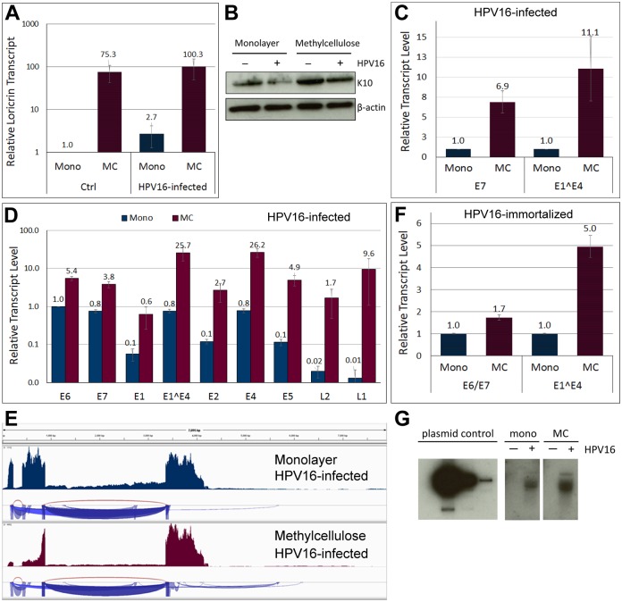
Fig 3. Differentiation activates both viral promoters and results in genome amplification.
HFKs maintained and infected in the presence of 10 μM Y-27632 for 5 days with HPV16 were subjected for 24–48 h to differentiation by plating in MC. (A) RT-qPCR analysis of loricrin transcript level in cells grown in monolayer and MC. The data shown are fold changes relative to uninfected HFK cells grown in monolayer. Error bars represent SEM of three independent experiments. (B) Western blot for keratin 10 in cells grown in monolayer and MC. (C) Expression of E7 and E1^E4 in HPV16 infected HFKs. The data shown are fold changes relative to cells grown in monolayer. Error bars represent SEM of three independent experiments. (D) Relative expression levels of individual ORFs in differentiated HPV16 infected cells. The data shown are fold changes normalized to E6 transcript levels in infected cells grown in monolayer. Error bars represent SEM of four biological replicates. (E) Read depths maps of viral transcripts isolated from HPV16-infected HFK grown in monolayer and MC. (F) Expression of E6/E7 and E1^E4 in HPV16 immortalized HFKs. The data shown are fold changes relative to cells grown in monolayer. Error bars represent SEM of six independent experiments. (G) Southern blot for viral genome isolated from HPV16-infected HFK grown in monolayer and MC.

