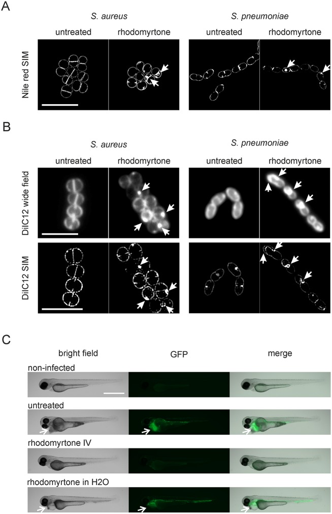
Fig 10. Effects of rhodomyrtone on S. aureus and S. pneumoniae and efficacy of rhodomyrtone against S. pneumoniae infection.
S. aureus RN4220 and S. pneumoniae D39 were grown until early log phase and treated with 1 μg/ml rhodomyrtone for 10 min. (A) SIM pictures of S. aureus and S. pneumoniae stained with Nile red. Arrows indicate membrane invaginations in rhodomyrtone-treated cells. Scale bar 2 μm. (B) DiIC12-stained cells show bright fluorescent patches after treatment with DiIC12 (arrows, upper panels). Corresponding SIM images show that these patches are membrane invaginations (arrows, lower panels). Scale bars 2 μm. (C) Rhodomyrtone reduces the bacterial load and reduces damage to the heart region in zebra fish embryos infected with S. pneumoniae. One day old zebra fish embryos were injected with 160 CFU of S. pneumoniae JWV500 expressing HlpA-GFP in the tail vein. Fish were treated with two injections (45 and 75 min post infection) of 25 ng rhodomyrtone each. For treatment in water, 5 μg/ml rhodomyrtone were directly added to the water. Pictures were taken 18 hours post infection. Note the visible damage to the heart region of the fish caused by S. pneumoniae infection (arrows, left panel) and the bright fluorescence spots, indicating the presence of S. pneumoniae JW500 in this region (arrows, middle panel). Reduction of the fluorescence signal compared to untreated cells was consistently observed in all rhodomyrtone-injected fish (see also S18 Fig), but not in fish treated by addition of the compound to the water. Experiments were performed in biological triplicates with 15 fish per condition in each replicate. Scale bar 200 μm.

