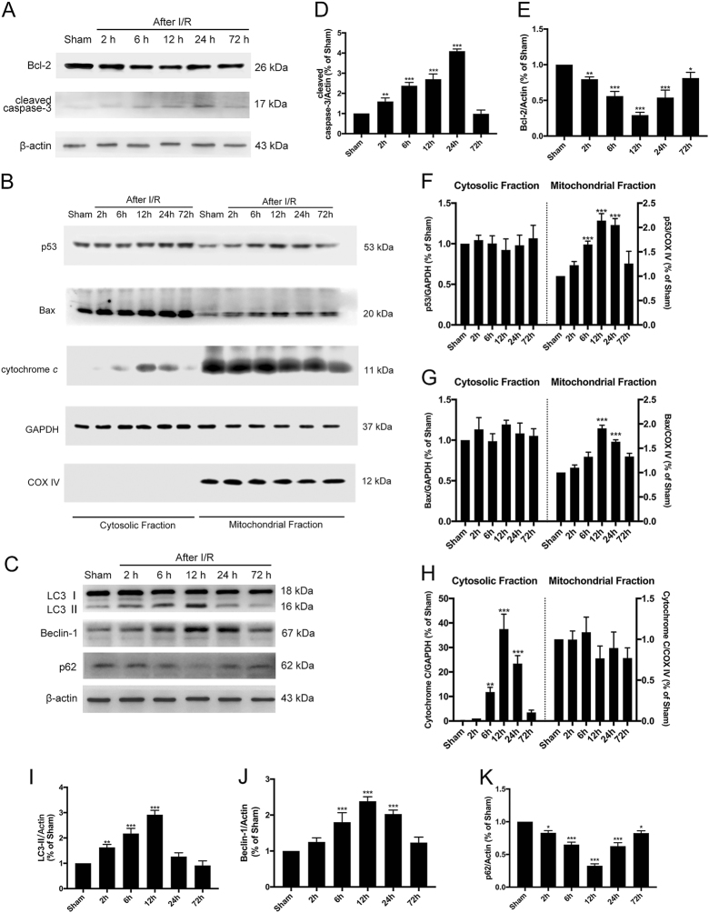
Fig. 1. Temporal changes of autophagy and apoptosis after tGCI.
a Immunoblots of the apoptosis-related proteins cleaved Caspase-3 and Bcl-2. Total cell lysates from the hippocampus were used, and β-actin was used as an internal control. b Immunoblots of p53, Bax, and cytochrome c in the cytosolic and mitochondrial fractions. GAPDH was used as a loading control for cytosolic proteins, and COX IV was used as the loading control for mitochondrial proteins. c Immunoblots of the autophagy-related proteins LC3B and Beclin-1. Total cell lysates from the hippocampus were used. d–k Densitometric analysis (mean ± SEM, n = 3 animals per group) of the proteins from (a, b, c) normalized to the respective loading controls. *P < 0.05 vs. sham + vehicle, **P < 0.01 vs. sham + vehicle, ***P < 0.001 vs. sham + vehicle

