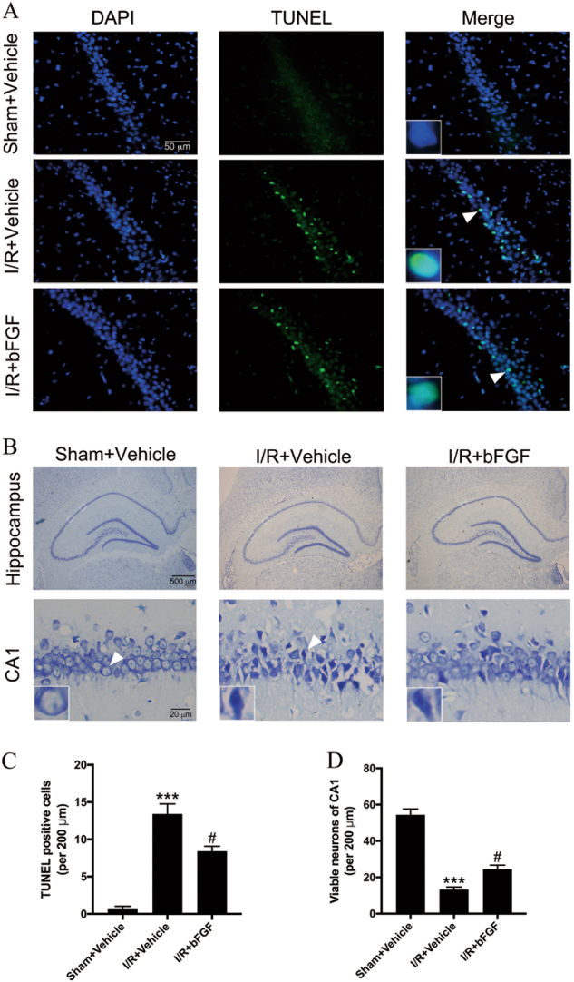
Fig. 2. bFGF treatment generated a significant neuroprotective effect in the hippocampal CA1 region after global cerebral I/R.

a Representative images of TUNEL staining in the CA1 region of the hippocampus 24 h after I/R. b Representative cresyl violet staining of the entire hippocampus and the detailed CA1 region 72 h after I/R. The white boxes in the hippocampus denote the area of the detailed CA1 region. Insets in the pictures are magnified neurons from the areas indicated by the arrowhead. c–d Quantitative data (mean ± SEM, n = 5–6 animals per group) of TUNEL-positive cells and viable neurons per 200-μm length of the medial CA1 region. ***P < 0.001 vs. sham + vehicle, #P < 0.05 vs. I/R + vehicle
