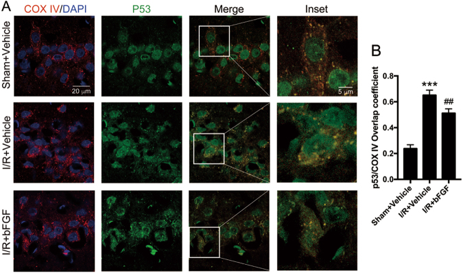
Fig. 7. bFGF administration inhibited the translocation of p53 to mitochondria.
a Representative confocal images of DAPI (blue), p53 (green), and COX IV (red) staining in the hippocampal CA1 region. COX IV was used as a mitochondrial marker, and nuclei were counterstained with DAPI. In the merged microphotographs, yellow puncta indicate the co-localization of p53 and mitochondria. b Manders’ overlap coefficient represents the quantification of co-localization of p53 and COX IV. At least ten cells from each group were selected randomly. ***P < 0.001 vs. sham + vehicle; ##P < 0.01 vs. I/R + vehicle group

