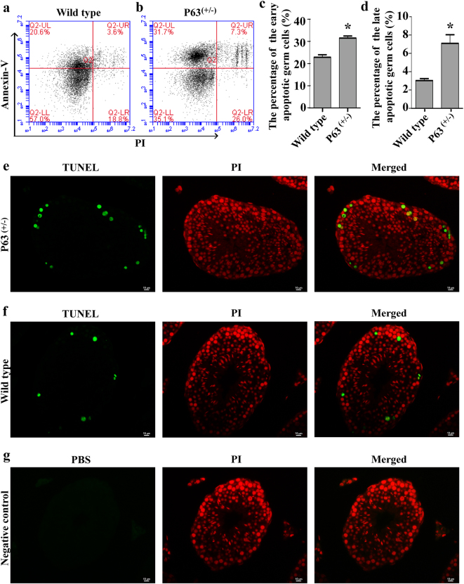
Fig. 4. The apoptosis in male germ cells of the P63(+/−) mice and wild-type mice.
The percentages of apoptotic male germ cells in wild-type mice a and P63(+/−) mice b at 2 months old. The percentages of early apoptosis c and late apoptosis d in male germ cells of wild-type mice and P63(+/−) mice were calculated using Student’s t-test. * indicated statistically significant differences (p < 0.05). TUNEL assay demonstrated the TUNEL-positive cells (green fluorescence) in P63(+/−) mice e and wild-type mice f. Replacement the TdT enzyme with PBS was used as the negative control g. PI (red fluorescence) was used to label cell nuclei. Scale bars in e–g = 10 μm. The data in Fig. 4a–g were presented from three independent experiments

