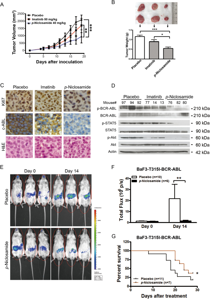
Fig. 6. p-Niclosamide abrogates the growth of T315I-BCR-ABL-harboring cells in immunodeficient mice and prolongs the survival of allograft mice.
a Nude mice bearing KBM5-T315I xenograft tumors were treated with placebo (PBS), imatinib (served as a control) or p-niclosamide. Tumor volumes were plotted against days post CML cell inoculation. ***P < 0.0001, one-way ANOVA, compared placebo-treated or imatinib-treated mice with p-niclosamide-treated ones at the last time point. Data are mean ± 95% confidence intervals. b Tumor weights were assessed after two-week of treatment (bottom) and representative tumors of each group are shown (top). *P < 0.05, ***P < 0.0001, compared with Placebo, one-way ANOVA, post hoc intergroup comparisons. c Immunohistochemical analysis of Ki67 and c-ABL in KBM5-T315I xenografted tissues from mice sacrificed 19 days after inoculation of tumor cells. Hematoxylin and eosin (H&E)-stained sections of the same tissues were shown. d Western blotting analysis of BCR-ABL and its target molecules in xenografted tissues from the mice treated with placebo, p-niclosamide or imatinib. e, f Niclosamide inhibited the proliferation of BaF3-T315I-BCR-ABL-Luc cells in NOD/SCID mice. BaF3-T315I-BCR-ABL-Luc cells were injected into mice via tail vein, allowed to grow for 5 days, and then treated with placebo or p-niclosamide for 2 weeks. Representative photos of mice after treatment (e) and bar chart of in vivo luminescence signals (f) were shown. *P < 0.05, Student’s t-test. g Effect of p-niclosamide on survival of mice bearing BaF3-T315I-BCR-ABL-Luc cells. Kaplan–Meier survival curve of CML mice after treatment of p-niclosamide. *P = 0.0346, Log-Rank test

