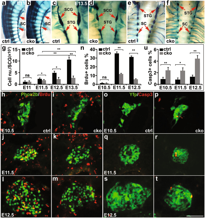
Fig. 1. Neural crest specific ablation of Isl1 leads to loss of sympathetic neurons.
a, b Wholemount X-gal staining revealed that initial formation of primary sympathetic ganglia appeared to be normal in Wnt1-Cre;Isl1f/f embryos (CKO) compared with Wnt1-Cre;Isl1+/+ (ctrl) littermates at E11. Scale bar, 500 µm. c–f From E11.5 onward, the size of the superior cervical ganglia (SCG), stellate ganglia (STG), and sympathetic chain (SC) of Isl1 CKO embryos became progressively reduced. Scale bar, 1 mm (c, d), 2 mm (e, f). g Quantitative analysis revealed a significant reduction in the total number of sympathetic neurons (X-gal+) per ganglion or sympathetic chain (E11) in Isl1 CKO embryos compared with control littermates at all developmental stages examined except E11. Error bars represent the s.d., n = 4, E11 (p = 0.54), E11.5 (p = 0.0183), E12.5 (p = 0.0106), E13.5 (p = 2e−05), two-tailed t-test. h–n BrdU labeling revealed a significantly reduced proliferation of Isl1 CKO sympathetic neurons (Phox2b+) at E11.5 and E12.5. At E10.5, no BrdU immunoreactivity was detected in both control and CKO mutant embryos. Error bars represent the s.d., n = 4, E10.5 (p = 0.594), E11.5 (p = 6.6e−06), E12.5 (p = 4.9e−08), two-tailed t-test. Scale bar, 50 µm. o–u Caspase-3 immunostaining showed significantly increased cell death in the SCG of Isl1 CKO embryos (on Rosa-YFP background) at E10.5, E11.5, and E12.5. Error bars represent the s.d., n = 4, E10.5 (p = 0.0016), E11.5 (p = 0.0103), E12.5 (p = 0.0002), two-tailed t-test. Scale bar, 50 µm. Asterisks (*) indicate statistically significant difference between the two groups indicated in brackets, ns = not significant, * p < 0.05, or ** p < 0.01

