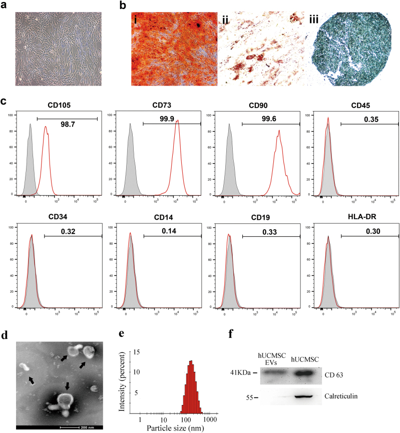
Fig. 1. Identification of human umbilical cord mesenchymal stem cells (hUCMSCs) and hUCMSC-derived extracellular vesicles (hUCMSC-EVs).
a The cell morphology of hUCMSCs (passage 3) was observed under a light microscope (magnification, ×100). b Representative images of osteocyte (×100), adipocyte (×400), and chondrocyte (×200) differentiation of hUCMSCs cultured in the differentiation media. The cells were analyzed using cytochemical staining with Alizarin Red (i), Oil red O (ii), and Alcian Blue (iii), respectively. c Flow cytometric analysis of the expression of cell surface markers related to MSCs. d Transmission electron microscopic images of hUCMSC-EVs. The scale bars indicate 200 nm, and the arrows indicate typical hUCMSC-EVs. e The size distribution of the hUCMSC-EVs was examined using a Zetasizer Nano ZS. f The positive marker for EVs, CD63, was detected in hUCMSC-EVs using western blotting, whereas the negative marker calreticulin was not detected

