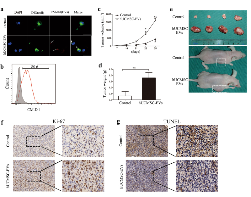
Fig. 3. Effects of hUCMSC-EVs pre-stimulation on tumor growth in vivo.
a DIO-labeled H1299 cells (green) were incubated with CM-Dil-labeled hUCMSC-EVs (red) for 2 h, and the hUCMSC-EV uptake by the LUAD cells was determined. The arrows indicate the fusion of the membrane. The scale bars indicate 50 μm. b FACS analysis of H1299 cells that had been treated for 12 h with CM-Dil-labeled hUCMSC-EVs or serum-free medium as control. The H1299 cells were pre-stimulated with hUCMSC-EVs for 12 h before they were injected into the nude mice. c The tumor volumes were measured, and d the tumor weights were determined at 35 days. e Imaging of tumors and tumor-bearing mice from the hUCMSC-EV-pre-stimulated groups at 35 days after the injections. f Ki-67 and g TUNEL immunohistochemical staining on tumor sections from the control or hUCMSC-EV-pre-stimulated group. The images are shown at ×100 (left panel) and ×400 (right panel)

