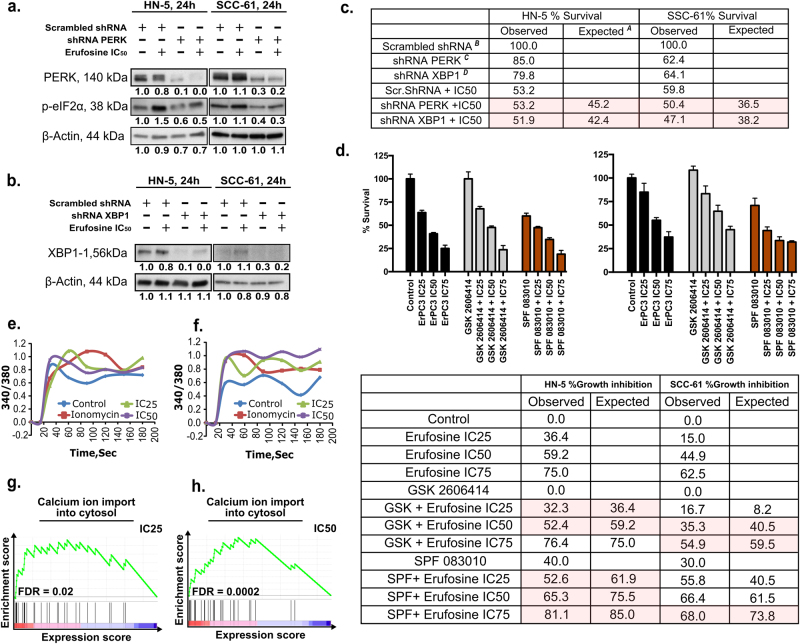
Fig. 3. UPR sensors contribute to erufosine’s antiproliferative activity.
a shRNA PERK knockdown in HN-5 and SCC-61 cells using lentivirus. Successful inhibition of PERK and its downstream mediator p-EIF2α are seen when compared to scrambled shRNA control. b Successful knockdown of XBP1 using lentivirus particle in HN-5 and SCC-61 cells. β-Actin was used as loading standard. c Percentage survival of scrambled, shPERK, and shXBP1 cells in response to erufosine in two cell lines. The expected survival is calculated by multiplying the individual survival percentages of each treatment arm (A = (B×C)/100 and A = (B×D)/100). d Percentage growth inhibition of wild-type cells in combination with erufosine and PERK inhibitor GSK-2606414 or IRE-1α inhibitor STF-083010. The expected growth inhibition is initially calculated as for survival under c and then by subtracting this value from 100. The highlighted columns show additive nature of combination at those doses. e, f Induction of Ca2+ release post erufosine treatment. Both cell lines showed an instantaneous increase in the intracellular Ca2+ levels as detected by Fura-2-AM dye post erufosine treatment. Ionomycin was used as positive control, and the signal was detected instantaneously post-drug exposure for 3 min. g, h Positively correlated enrichment of the term “calcium ion import in cytosol” upon erufosine exposure in HN-5 cells at IC25 and IC50 concentrations.

