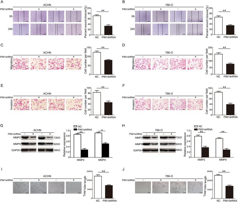
Fig. 3. Depletion of PIM1 inhibits the migration, invasion and angiogenesis behaviours of ccRCC cells in vitro.
a, b Wound-healing assays. The wound closure rate was calculated. Scale bar: 200 μm. c, d Transwell migration assays. The cell number per field was calculated. Scale bar: 20 μm. e, f Matrigel Transwell assays. The cell number per field was determined. Scale bar: 20 μm. g, h Immunoblotting analysis of MMP2 and MMP9 protein levels. GAPDH was used as the loading control, and the indicated proteins were quantified with ImageJ software. i, j Capillary tube formation (CTF) assays. The total length of tubes formed by HUVECs was calculated with ImageJ software. Scale bar: 50 μm. All data represent the mean ± S.E.M. of the values from three biological replicates. Statistical significance was determined with a two-tailed Student’s t-test. *P < 0.05; and **P < 0.01

