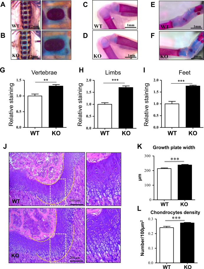
Fig. 5. PP5 deficiency increases growth plate thickness and enhances cartilage development.
Whole-mount Alcian blue and Alizarin red staining of WT and PP5 KO newborn mice showed stronger blue staining in vertebrae (a and b), limbs (c and d), and feet (e and f) in the PP5 KO. Alizarin red staining was quantified by ImageJ analysis (g–i). H&E staining of femurs revealed the area of growth plate in the KO mice was wider than that of WT mice (j). Quantitative analyses indicated the significant increase in growth plate width (k) and the density of chondrocytes (cell number/100 µm2) within the growth plate in KO mice (l). Magnified areas are indicated by dashed boxes. Values are presented as mean ± SE. **P < 0.01 and ***P < 0.005 using Student’s t-test. Each genotype, three same positions were selected from individual H&E staining of total three animals

