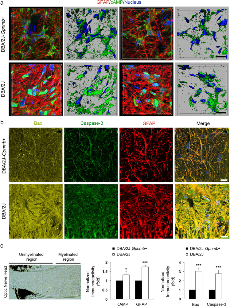
Fig. 5. Increased expression of cAMP as well as activated Bax and caspase-3 in astrocytes of the glial lamina in glaucomatous DBA2/J mice.
a and b Immunohistochemical analyses of cAMP (green), activated Bax (yellow), and caspase-3 (green) as well as GFAP (red) immunoreactivity in the ONH astrocytes in the glial lamina of age-matched non-glaucomatous DBA/2J-Gpnmb+ and 10-month-old glaucomatous DBA/2 J mice. Note that representative images showed increases of cAMP, activated Bax, and caspase-3 immunoreactivity in astrocytes of the glial lamina in glaucomatous DBA/2 J mice compared with non-glaucomatous DBA/2J-Gpnmb+. Nuclei were stained with Hoechst 33342. Scale bar, 20 μm. c Quantitative analysis showed significant increases of cAMP and GFAP, as well as activated Bax and caspase-3 immunoreactivity in glaucomatous DBA/2 J mice. For each determination, the immunoreactivity in controls was normalized to a value of 1.0. Data are shown as the mean ± S.D. (n = 5). *P < 0.05; ***P < 0.001 (two-tailed unpaired Student’s t-test)

