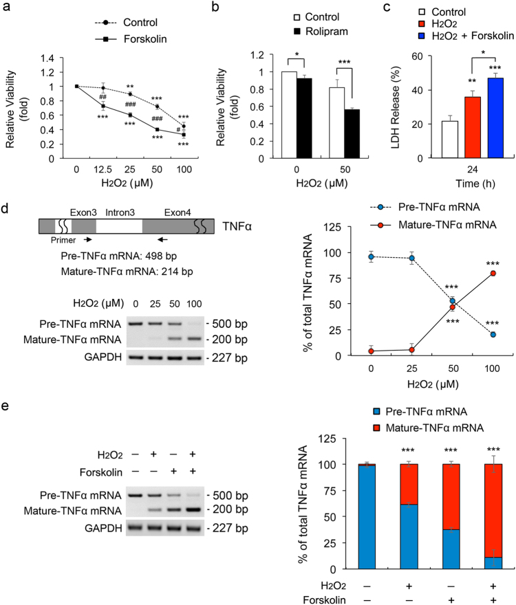
Fig. 6. Elevated intracellular cAMP exacerbates vulnerability to oxidative stress in ONH astrocytes.
a Cell viability assay in ONH astrocytes treated with H2O2 (50 μM) and/or forskolin (10 μM) for 1 h. b Cell viability analysis using MTT assay in ONH astrocytes treated with H2O2 (50 μM) and/or rolipram (2 μM) for 1 h. c Cell death analysis using LDH assay in ONH astrocytes treated with H2O2 (50 μM) and/or forskolin (10 μM) for 24 h. d Schematic illustration of rat pre-TNFα mRNA structure for RT-PCR analysis. The primer location was indicated by arrows. RT-PCR analysis of TNF-α splicing in ONH astrocytes treated with H2O2 (0–100 μM) for 1 h. e RT-PCR analysis of TNF-α splicing in ONH astrocytes treated with H2O2 (50 μM) and/or forskolin (10 μM) for 1 h. Data were normalized by GAPDH expression. For each determination, the cell viability, cell death and mRNA expression in controls was normalized to a value of 1.0 or 100%. Data are shown as the mean±S.D. (n = 3). *P < 0.05; **P < 0.01; ***P < 0.001 (two-tailed unpaired Student’s t-test)

