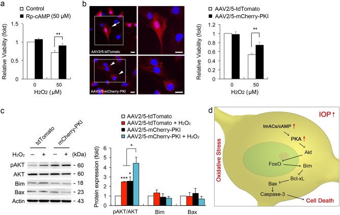
Fig. 8. PKA inhibition protects ONH astrocytes against oxidative stress.
a Cell viability analyses using MTT assay in H2O2 (50 μM for 1 h)-induced ONH astrocytes treated with Rp-cAMP (50 μM). b Cell viability analysis using MTT assay in H2O2 (50 μM)-induced ONH astrocytes transduced with AAV2/5-tdTomato or mCherry-PKI. Representative images showed tdTomato (red) or mCherry-PKI (red) expression in ONH astrocytes. Nuclei (blue) were stained by Hoechst 33342. Note that PKI overexpression promoted ONH astrocyte survival. Scale bars, 20 μm. c Immunoblot analyses of pAKT, total AKT, Bim and activated Bax in H2O2 (50 μM for 1 h)-induced ONH astrocytes transduced with AAV2/5-tdTomato or mCherry-PKI. d A hypothetical model for the role of tmAC activation-mediated cAMP/PKA signaling pathway in ONH astrocytes against glaucomatous insults such as elevated IOP and oxidative stress. For each determination, the cell viability as well as mRNA and protein expression in controls were normalized to a value of 1.0. Data are shown as the mean ± S.D. (n = 3). *P < 0.05; **P < 0.01; ***P < 0.001 (two-tailed unpaired Student’s t-test)

