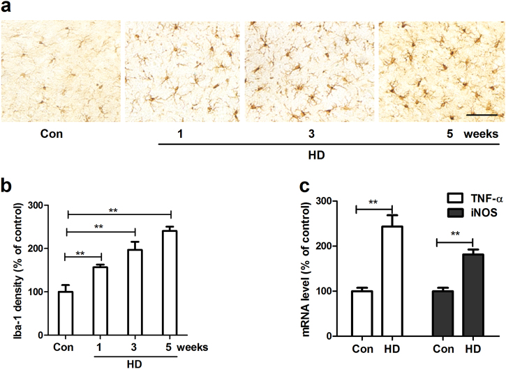
Fig. 2. HD exposure induces microglial activation in the SN of rats.
a After 1, 3, and 5 weeks of HD treatment, microglia in the SN were stained with antibody against Iba-1 and the representative images were shown. Activated microglia display larger cell body size and intensified Iba-1 staining. b Microglial activation was quantified by calculating the density Iba-1 immunostaining. c The gene expressions of TNF-α and iNOS were measured in the midbrain of HD-treated rats by using RT-PCR. *p < 0.05, **p < 0.01; n = 4–6; Scale bar = 50 μm

