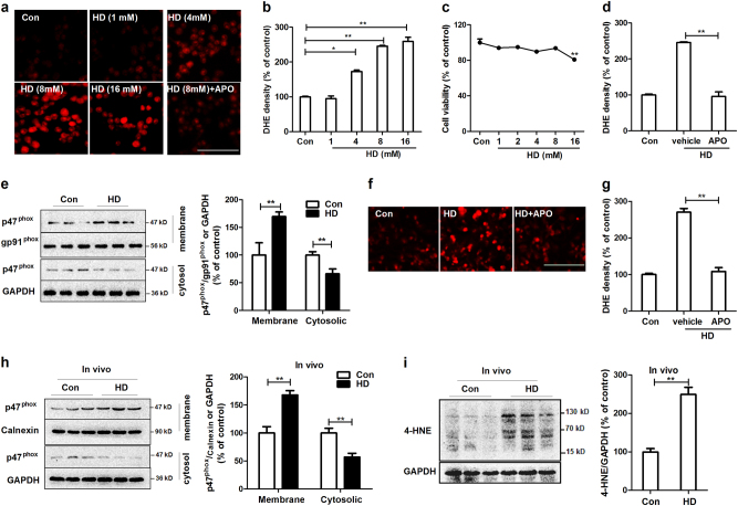
Fig. 4. HD activates NOX2.
a BV2 microglial cells were treated with 1, 4, 8, and 16 mM HD with or without apocynin (APO) pre-treatment. The production of superoxide was assessed by DHE and the representative images of DHE oxidation were shown. b The density of red fluorescence of DHE oxidation was quantified. c Cell viability of BV2 microglia treated with different concentrations of HD was detected using LDH assay. d The effects of apocynin on HD-induced superoxide production was quantified. e The membrane translocation of NOX2 cytosolic subunit, p47phox in HD-treated BV2 microglial cells was detected after 30 min of HD stimulation by using Western blot and the density of blots was quantified. Gp91phox, an abundant membrane protein, and GAPDH were used as an internal membrane and cytosolic control, respectively. Previous reports indicated that gp91phox is a reliable internal membrane control52,60. f HD-induced superoxide production was examined in primary mixed-glia cells by DHE and the representative images of DHE oxidation were shown. g The density of red fluorescence of DHE oxidation was quantified. Results were expressed as a percentage of controls from three experiments performed in duplicate. h The membrane translocation p47phox was detected in midbrain tissues of HD-treated rats by Western blot and the density of blots was quantified. i The levels of 4-HNE were determined in midbrains of HD-treated rats by Western blot and the density of blots was quantified. *p < 0.05, **p < 0.01; n = 3–6; Scale bar = 100 μm

