
Fig. 7. AP2M1 expression following apoptosis induction.
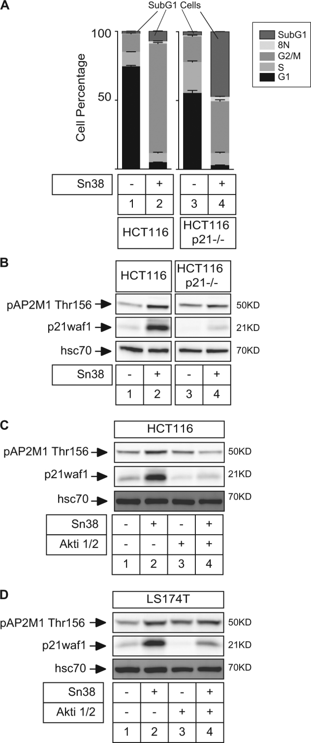
a HCT116 and HCT116 p21−/− cells have been treated with sn38 for 72 h (5 ng/ml) and apoptosis was evaluated by FACS analysis and the detection of subG1 cells (n = 3 ± s.d.). b Phospho-AP2M1 and p21waf1 expressions have been analyzed by western blot in the indicated cells (n = 3). c, d LS174T or HCT116 cells were treated with sn38 in the presence or absence of Akti 1/2 for 72 h (10 µM) and phospho-AP2M1 and p21waf1 expressions were analyzed as described above (n = 3)