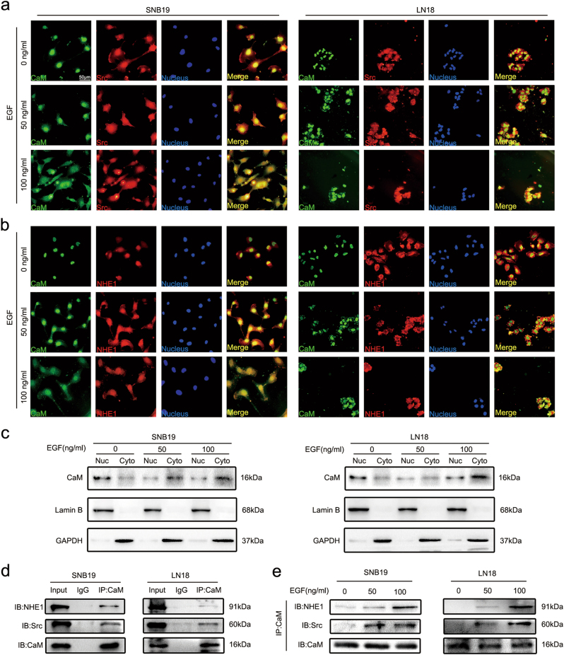
Fig. 6. EGF potentiated CaM redistribution from nucleus to cytoplasm and binding with Src and NHE1.
a EGF-induced CaM colocalization with Src in the cytoplasma by Immunofluorescence analysis. Green: CaM, Red: Src, Blue: Nucleus. (Representative images were presented. Images were taken from three different microscopic visions from every independent experiment). b EGF-induced CaM colocalization with NHE1 in the cytoplasma by Immunofluorescence analysis. Green: CaM, Red: Src, Blue: Nucleus. (Representative images were presented. Images were taken from three different microscopic visions from every independent experiment). c The distribution of CaM in nucleus and cytoplasm with different concentrations of EGF were detected by western blot. Lamin B1 and GAPDH served as nucleus (Nuc) and cytoplasmic (Cyto) loading control, respectively. d The combination between CaM and Src/NHE1 was detected by using co-immunoprecipitation assay without treatment with EGF. e Upon treatments with different concentrations of EGF, the binding of CaM with Src and NHE1 was explored by co-immunopreciptation

