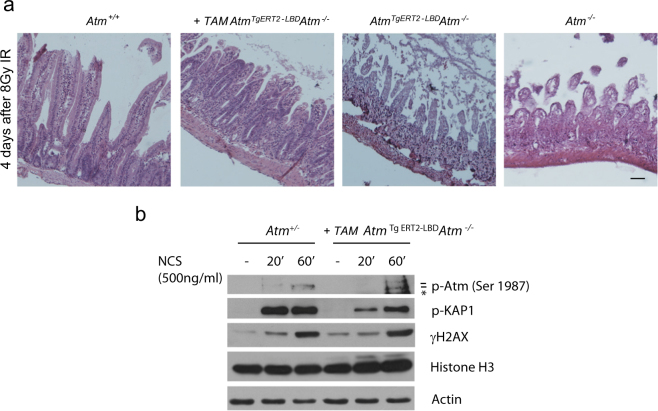
Fig. 4. DNA damage signaling reactivation in tamoxifen-treated AtmTgERT2-LBDAtm−/− mice.
a Representative image of small intestine sections stained with H&E. Mice were irradiated with 8 Gy and sacrificed 4 days after treatment. Scale bar = 100 μm. b Western blot of thymocytes collected from Atm+/− and AtmTgERT2-LBDAtm−/− mice 26 days after tamoxifen treatment. Cells were freshly isolated and exposed to NCS. Atm, Kap1, and H2AX phosphorylation are shown. Asterisk indicates a not specific band. Data are representative of two independent experiments

