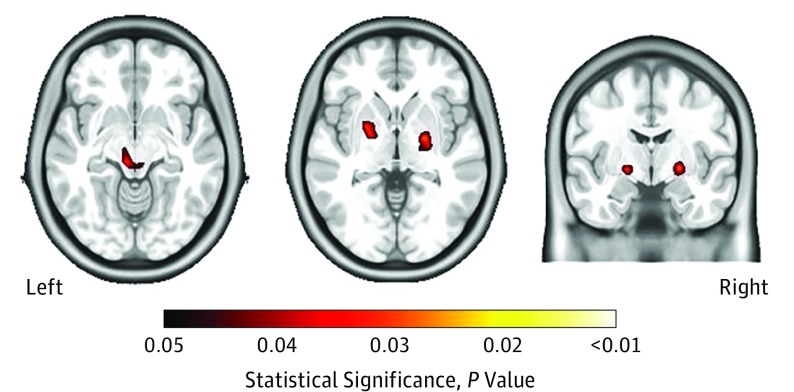
Figure 3. Smaller Subcortical Brain Regions and Lower Nadir CD4 Counts.
Smaller brain volumes in the putamen, globus pallidus, and thalamus, as revealed with tensor-based morphometry, correlated with lower nadir CD4 cell counts. The left image shows the thalamus, the center image shows the putamen and globus pallidus, and the right image shows the putamen and globus pallidus.

