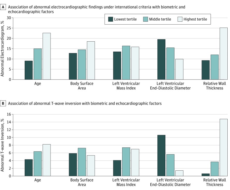
Figure. Association of Electrocardiographic Abnormalities With Biometric and Echocardiographic Parameters.
A, The prevalence of abnormal findings in the highest and lowest age tertiles differed significantly under international electrocardiographic interpretation criteria (P < .001), as did the highest and lowest relative wall thickness tertiles (P < .001); full statistics, including additional significant differences, are available in the Supplement. (Similar results were obtained using the Seattle and refined criteria.) B, The prevalence of T-wave inversion in the highest and lowest left ventricular end-diastolic diameter tertiles differed significantly (P < .001), as did the highest and lowest relative wall thickness tertiles (P < .001); full statistics, including significant differences, are available in the Supplement.

