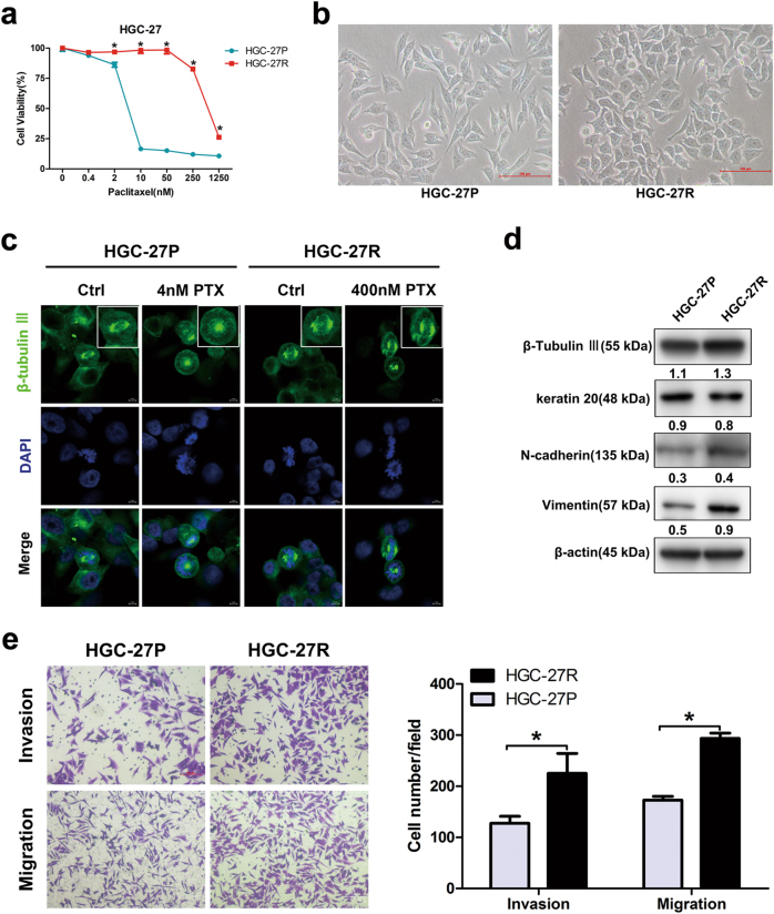
Fig. 1. Altered morphology, microtubular disorders, and EMT in PTX-resistant GC cells.
a Cell viability of HGC-27P and HGC-27R cells after exposure to PTX for 48 h. b Representative morphology of parental and PTX-resistant GC cells. Original magnification: ×200. c Microtubules at metaphase visualized with immunofluorescence after an exposure to PTX for 12 h. β-Tubulin III stained for microtubules and DAPI stained for nuclei (green and blue, respectively). Scale bar, 5 μm. d β-Tubulin III and EMT protein expression measured using western blot. e Invasion and migration capacity of HGC-27P and HGC-27R cells measured by Transwell assay with/without Matrigel. Data expressed as mean ± S.D. of three independent experiments. *p < 0.05

