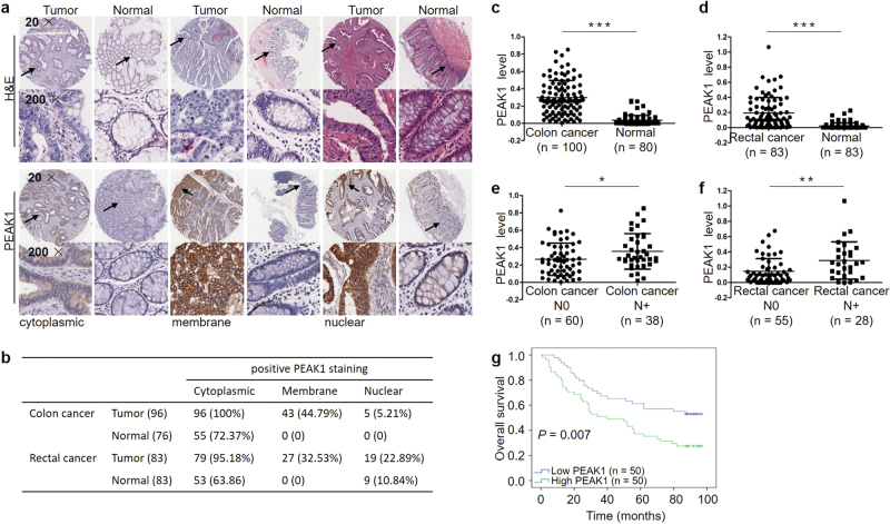
Fig. 1. PEAK1 protein expression levels in CRC specimens and prognostic significance.
a Representative immunohistochemical images of cytoplasmic, membrane and nuclear staining. b Summary of PEAK1-positive staining data for colon cancer and rectal cancer. c PEAK1 protein expression in 100 colon cancer and 80 normal tissues. Statistical significance was determined by a two-tailed, unpaired Student’s t-test. d PEAK1 protein expression in 83 pairs of rectal cancer. Statistical significance was determined by a two-tailed, paired Student’s t-test. e, f PEAK1 protein expression in colon cancer tissues without lymph node metastasis (N = 60) and with lymph node metastasis (N = 38) and rectal cancer tissues without lymph node metastasis (N = 55) and with lymph node metastasis (N = 28). Statistical significance was determined using a two-tailed, unpaired Student’s t-test. g Kaplan–Meier analysis of overall survival according to low and high PEAK1 protein expression in 100 colon cancer patients. (*P < 0.05, **P < 0.01, ***P < 0.001)

