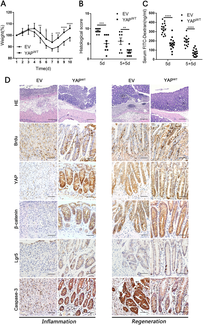
Fig. 4. YAP overexpression protected epithelial cells against DSS-induced colitis and promoted intestinal regeneration in mice.
(a) Body weight curves of YAPWT and EV mice during the successive 5-d DSS treatment, followed by 5 days of normal drinking water. Mice were killed on 5d (n = 9 each) and 5 + 5d (EV n = 8, YAPWT n = 9). Data represent the means ± SDs. (b) Histological scores in YAPWT and EV mice on 5d (n = 9 each) and 5 + 5d (EV n = 8, YAPWT n = 9). Data represent the means ± SEM. **P < 0.01, ***P < 0.001. (c) Serum FITC-dextran in YAPWT and EV mice on 5d (n = 15 each) and 5 + 5d (EV n = 14, YAPWT n = 15). Data represent the means ± SEM. ****P < 0.0001. (d) Paraffin-embedded sections of YAPWT and EV mice colons were analysed by H&E (× 100) and immunohistochemical staining (× 400) at 5d (Left: inflammation) and 5 + 5d (Right: regeneration). Scale bars = 200 μm

