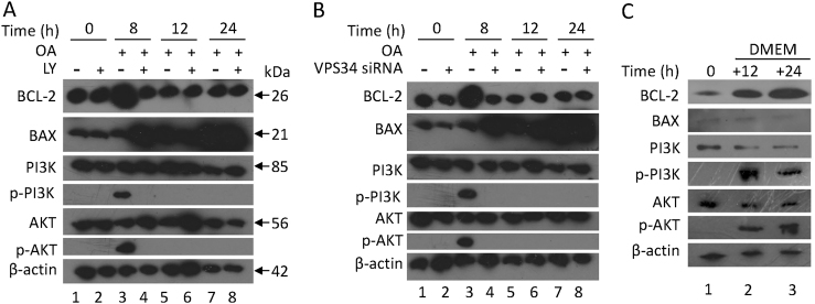
Fig. 5. OA treatment activates PI3K/AKT signaling pathway and increases BCL-2 expression at 8 h, whereas increases BAX expression at 24 h.
HepG2 cells were pre-treated with LY294002 (a) or VPS34 siRNA (b) and then were cultured with 400 μM OA. c HepG2 cells were treated with 400 μM OA for 8 h and then were cultured in normal DMEM culture for 12 and 24 h. a–c Lysates of HepG2 cells were collected, and an immunoblot assay was conducted with the indicated antibodies

