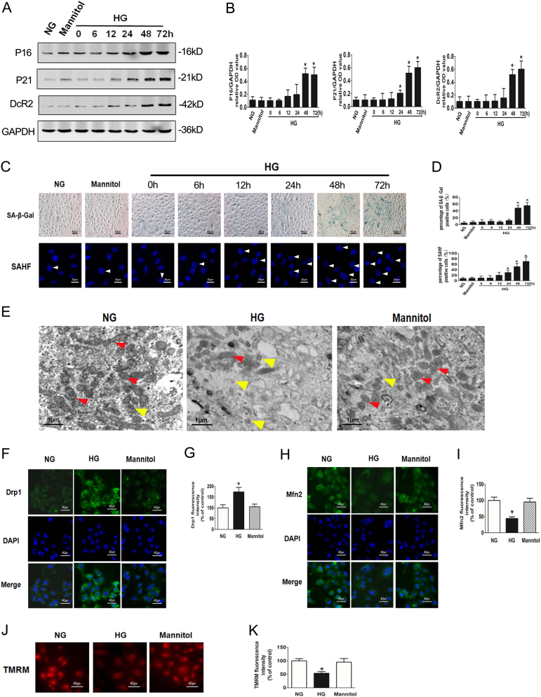
Fig. 1. HG induces premature senescence and mitochondrial dysfunction in mouse RTECs.
a Western blot analysis of P16, P21, DcR2 expression in mouse RTECs treated with NG, Mannitol, or HG for 0–72 h. b Densitometry of the respective blots from three independent experiments. *P < 0.05 vs. HG 0 h. c SA-β-gal (upper row) and SAHF (lower row) were detected in mouse RTECs. The white arrows indicate the nucleus of SAHF. d Percentage of SA-β-gal (upper row) or SAHF (lower row)-positive RTECs. e Electron microscopy images of RTECs showing changes in mitochondrial morphology under HG condition for 48 h. The red arrows indicate normal mitochondria, while the yellow arrows indicate mitochondrial fragments. f Drp1 expression in RTECs after HG stimulation for 48 h. g Analysis of Drp1 fluorescence intensity. *P < 0.05 vs. NG. h Mfn2 expression in RTECs after HG stimulation for 48 h. i Analysis of Mfn2 fluorescence intensity. j Mitochondrial membrane potential was measured using TMRM fluorescence. k Analysis of TMRM fluorescence intensity

