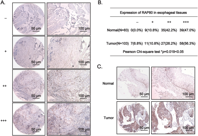
Fig. 1. RAP80 is highly overexpressed in esophageal squamous tumor tissues.
a The criteria for defining the expression level of RAP80 in the tissue microarray (TMA), detected by IHC staining, based on the percentage of cells with different staining intensities at two magnifications (50 and 100 μm). b The Pearson χ2 test of RAP80 in esophageal normal tissues (n = 83) and tumor tissues (n = 103). *p = 0.019. c The representative pictures of RAP80 staining in TMA. Scale bar, 50 and 100 μm, respectively

