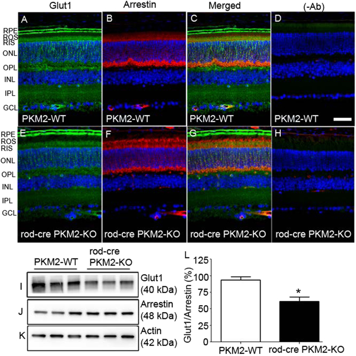
Fig. 12. Effect of PKM2 loss on glucose transporter Glut1 expression in the retina.
Prefer-fixed sections of wild-type a- d and rod-cre PKM2-KO e–h mouse retinas were subjected to immunofluorescence with anti-Glut1 a, c, e, g and anti-arrestin b, c, f, g antibodies. c, g Merged images of Glut1 and arrestin. d, h Omission of primary antibodies. GCL, ganglion cell layer; INL, inner nuclear layer; IPL, inner plexiform layer; ONL, outer nuclear layer; OPL, outer plexiform layer; RIS, rod inner segments; RPE, retinal pigment epithelium; ROS, rod outer segments. Scale bar = 50 μm. Retinal proteins prepared from three independent wild-type and rod-cre PKM2-KO mice were subjected to immunoblot analysis with anti-Glut1 i, anti-arrestin j, and anti-actin k antibodies. Densitometric analysis of immunoblots was performed in the linear range of detection. Absolute values were then normalized to arrestin l. Data are mean ± SEM (n = 3). *p < 0.01. The normalized PKM2 wild-type control was set as 100%

