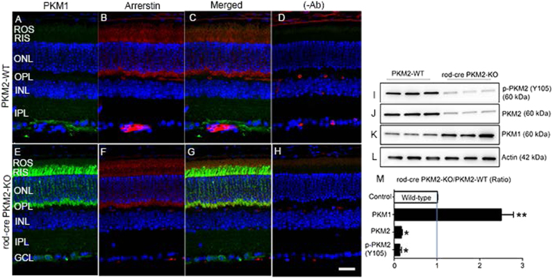
Fig. 2. Immunofluorescence analysis of PKM1 in wild-type and rod-cre PKM2-KO mice.
Prefer-fixed sections of wild-type a- d and rod-cre PKM2-KO e–h) mouse retinas were subjected to immunofluorescence with anti-PKM1 a, e and anti-arrestin b, f antibodies. c, g Merged images of PKM1 and arrestin. d, h Omission of primary antibodies. GCL, ganglion cell layer; INL, inner nuclear layer; IPL, inner plexiform layer; ONL, outer nuclear layer; OPL, outer plexiform layer; RIS, rod inner segment; ROS, rod outer segments. Scale bar = 50 μm. Retinal homogenates (5.0 µg protein) from wild-type and rod-cre PKM2 KO mice were subjected to immunoblot analysis with anti-pPKM2 (Y105) i, anti-PKM2 j, anti-PKM1 k, and anti-actin l antibodies. We normalized the protein expression/phosphorylation to actin m and then calculated the ratios (rod-cre PKM2-KO/PKM2-WT). Data are mean ± SEM (n = 3). *p < 0.004, **p < 0.001

