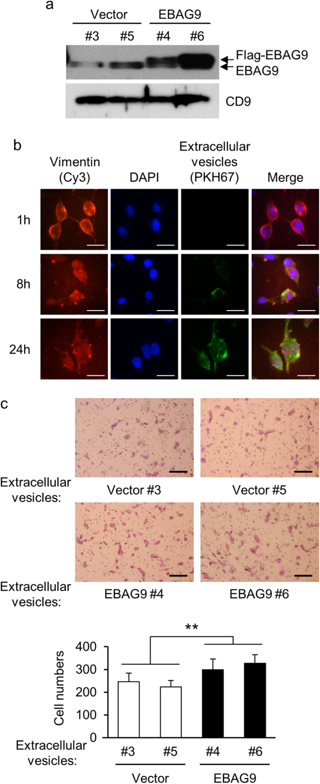
Fig. 5. Extracellular vesicle (EV)-transferred EBAG9 stimulates cancer cell migration.

a EBAG9 protein is secreted in EVs. EVs were prepared from LNCaP-EBAG9 cells (EBAG9 #4 and #6) and control cells (Vector #3 and #5), and subjected to western blot analysis using EBAG9 antibody. b EVs are integrated into LNCaP cells. Cells were co-cultured with green fluorescent PKH67-labeled EVs for indicated times and co-stained with Cy3-labeled anti-vimentin antibody and 4′,6-diamidino-2-phenylindole (DAPI). Scale bars, 10 μm. c Cancer-derived EVs containing EBAG9 stimulate parental cell migration. LNCaP cells were incubated with LNCaP-EBAG9 cell-derived (EBAG9 #4 and #6) or control cell-derived (Vector #3 and #5) EVs, and cell migration was evaluated as described in Fig. 4. Data are shown as mean ± SD (n = 5). **P < 0.01 (Statistical analysis was performed using the two-sided Mann–Whitney U-test)
