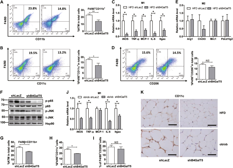
Fig. 6. Downregulation of B4GalT5 reduce HFD-induced M1 macrophage infiltration in subcutaneous adipose tissue.
Six-week-old C57BL/6 J mice and ob/ob mice were injected with adenovirus expressing B4GalT5 or control LacZ shRNA twice a week s.c. adjacent to both sides of the inguinal fat pads for 6 weeks. HFD feeding started at 6 weeks of age on WT mice. a–f HFD mice; g–j ob/ob mice. a The adipose macrophages (CD11b+F4/80+) were examined by flow cytometry analysis from SVF. b, d FACS analysis determined iWAT F4/80+CD11c+M1and F4/80+CD206+ M2 macrophages (n = 6/group). c, e qPCR determined M1 markers, iNOS, TNF-α, MCP-1, IL-6, Itgax, and M2 markers, Arg1, Chi3l3, Mrc1, Pdec1lg2 in iWAT from different mouse group as indicated (n = 6/group). f Western blotting analysis of the p-JNK, total(t)-JNK, p-p65, and p65 in the iWAT. g–i FACS analysis determined iWAT total macrophages (CD11b+F4/80+), F4/80+CD11c+M1and F4/80+CD206+ M2 macrophages. j mRNA level of M1 markers. k CD11c IHC of iWAT sections (five images per mouse), scale bars 20 µm. Each data is representative of at least three independent experiments with identical results. *P < 0.05, **P < 0.01, ***P < 0.001

