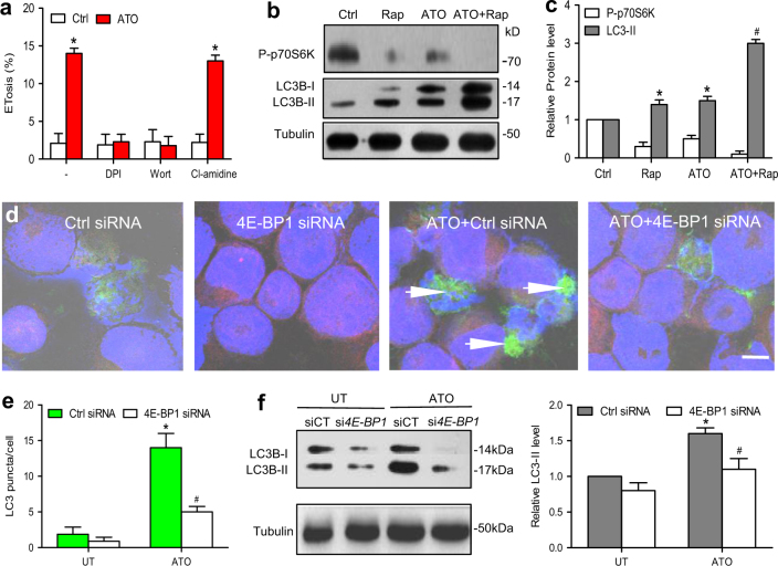
Fig. 3. ATO induce autophagy through the mTOR pathway.
a NB4 cells were preincubated with the indicated inhibitors (pretreatment with DPI (10 μM, 4 h), wortmannin (1 mg/ml) for 30 min, Cl-amidine (200 μM)) before treatment with ATO (0.75 μM, 48 h) to induce ETosis. b, c NB4 cells were treated with vehicle or rapamycin (10 nM) in the presence or absence of ATO (0.75 μM) for 48 h. Cell lysates were examined by western blot by the use of anti-P-p70S6K, anti-LC3, or anti-tubulin antibodies. Quantification of the data shown in panel c (n = 3). *P < 0.05 vs. control. d NB4 cells were stained with DAPI (blue) and anti-LC3 (green). Immunofluorescence images showed inhibition of the negative downstream effector of mTOR pathway by knocking down 4E-BP1 reduced the aggregation of LC3 (green). e The LC3 puncta per cell in ATO-treated NB4 cells measured by immunofluorescence (n = 3). f The levels of LC3B-I and LC3B-II were detected by western blotting (n = 3). All values are mean ± SD. *P < 0.05 vs. untreated group, #P < 0.05 vs. control e, f. Bars represent 20 μm in d. DPI diphenyleneiodonium chloride, Wort Wortmannin, Rap rapamycin (mTOR inhibitor), PAD4 peptidylarginine deiminase 4, UT untreated group, Ctrl, control

