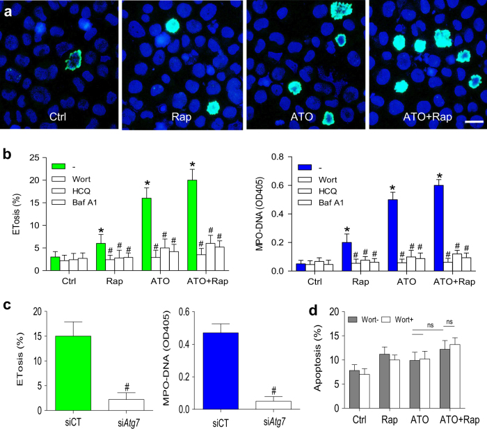
Fig. 4. Rapamycin enhances the effects of ATO on autophagy-dependent ETosis.
a, b NB4 cells were cultured in rapamycin (10 nM) or/and ATO (0.75 μM) for 48 h and treated with vehicle or with various autophagy inhibitors (wortmannin, HCQ and Baf A1). a NB4 cells were stained with DAPI (blue) and anti-histone-3 (green). Immunostaining images showed ATO-induced ETosis was enhanced by concomitant addition of rapamycin. b Quantification of the percentage of ETotic cells and corresponding MPO–DNA complexes concentrations for the different treatments (n = 5). c NB4 cells were transiently transfected with Atg7 siRNA (siAtg7) at a concentration of 100 nM and scrambled siRNA (scr) was used as a negative control (siCT). Seventy-two hours after transfection, cells were treated with ATO (0.75 μM) for 48 h. The percentage of ETosis and the concentration of MPO–DNA complexes were then measured (n = 5). d Apoptosis was analyzed by flow cytometry (n = 5). All values are mean ± SD. *P < 0.05 vs. control, #P < 0.05 vs. Wort-, ns = not significant. Bars represent 20 μm in a. UT untreated group, Rap rapamycin, Wort wortmannin

