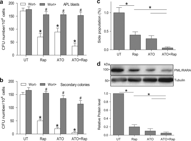
Fig. 6. ETosis contributes to LICs loss in APL in vitro.
a Primary APL cells isolated from patients were cultured in methylcellulose for a week with rapamycin (10 nM) or ATO (0.75 μM) alone or in combination and in the presence or absence of wortmannin (1 mg/ml). Number of colonies is shown (n = 8). b After 1 week of treatment, cells were harvested, washed and cultured in semisolid medium in the absence of any drug (n = 8). Number of secondary colonies is shown. *P < 0.05 vs. UT, #P < 0.05 vs. wortmannin (-). c, d NB4 cells treated with rapamycin (10 nM) and/or ATO (0.75 μM) for 48 h. c NB4 side population was defined by exclusion of Hoechst 33342 in the absence or presence of verapamil. The percentage of cells representing the side population was measured. d Lysates from NB4 cells were analyzed by immunoblotting by the use of antibodies against anti-PML/RARA, and tubulin was used as a loading control (n = 5), *P < 0.05. Wort, wortmannin

