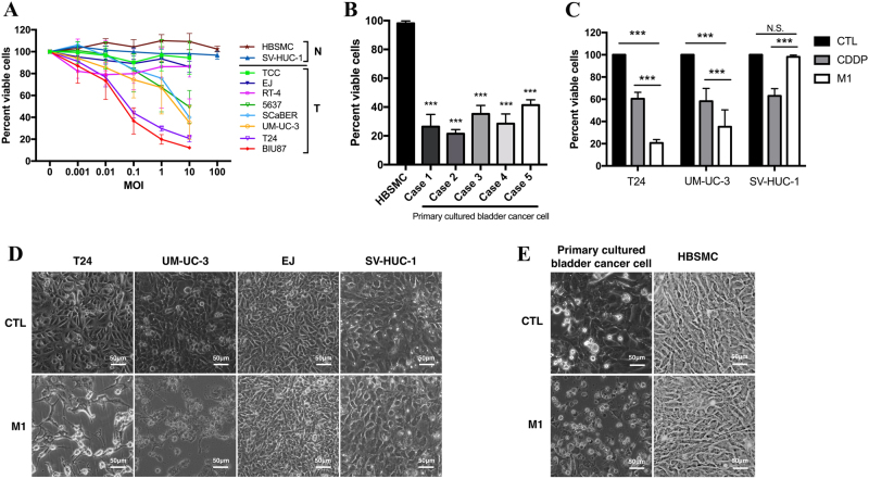
Fig. 1. Effect of M1 on bladder cancer cell viability.
a Evaluations of the viability of several bladder tumor (T) and normal (N) cell lines were performed by MTT assay after the cells were exposed to M1. Each color represents one cell line. b Five primary cultured bladder cancer tissue specimens were isolated from a clinical bladder cancer tumor specimen, and the responsiveness of each specimen to M1 was assessed and compared with that of a primary normal bladder tissue specimen (HBSMCs). c The bladder cancer cell lines T24 and UM-UC-3 and the normal cell line SV-HUC-1 were treated with CDDP (10 μM) and M1 (MOI = 10 PFU per cell), and cell viability assay was performed at 48 h post-infection. d, e Observation of the morphology of various bladder cells exposed to M1 (MOI = 10 PFU per cell) for 48 h. d Bladder cancer cells (T24, UM-UC-3, and EJ) and normal cells (SV-HUC-1), e Primary cultured bladder cancer cells and normal cells (HBSMCs). ***p < 0.001; N.S., not significant

