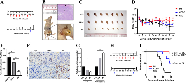
Fig. 5. M1 significantly reduces orthotopic bladder tumor sizes.
Establishment of the orthotopic bladder cancer mouse model. Laparotomy and bladder wall injections was used to establish the animal model. a We divided the animals into control, CDDP and M1 group (seven mice in each group), treatment in each group: CDDP, intraperitoneally at 2 mg/kg two times daily; M1, intravenously at 8.7 × 10⁷ PFU once daily for 8 days; and control, sterile PBS intravenously once daily for 8 days. i.v. intravenous injection, i.p. intraperitoneal injection. b Tumors visible to the naked eye formed 5–7 days after tumor inoculation. The image in the bottom-right corner is a high-magnification image of the image on the left. Arrow, bladder tumor; arrowhead, normal bladder smooth muscle; star, filled bladder cavity. c At the end of the experiment, all the mice were anesthetized and euthanized, and their tumor-bearing bladders were harvested and photographed. d The body weights of the tumor-bearing mice were recorded. e The weights of the tumor-bearing bladders were measured and recorded. f Immunohistochemistry was performed to detect Ki-67 and cleaved-caspase-3 expression. g Ki-67 and cleaved-caspase-3 expression levels in treated cells were compared with those in control cells, Relative protein expression levels were quantified with Image-Pro Plus 6.0 (IPP 6.0, Media Cybernetics, Rockville, MD). CTL control. h Timeline of experiment setup for i. Kaplan–Meier survival curve of mice bearing UM-UC-3 orthotopic tumors treated with PBS, CDDP, and M1 virus. Log‐rank with Holm–Sidak multiple comparison. *p < 0.05; **p < 0.01; ***p < 0.001

