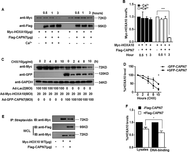
Fig. 4. CAPN7-mediated HOXA10 degradation impairs the protein stability and DNA-binding ability of HOXA10.
a CAPN7 degrades HOXA10 in vitro. In the presence of Ca2+ (3 mM) for 3 h at 37 °C. Lysates were analyzed by western blotting with the indicated antibodies. b The intensities of Myc-HOXA10 signals were quantified from the different groups and normalized to the control group (hour = 0 h). Values represent the mean ± SEM (n = 3), ***p < 0.001 vs hour = 0 group. c Ishikawa cells were transduced with Ad-GFP-CAPN7 (100 MOI) and Ad-Myc-HOXA10 or with Ad-Myc-HOXA10 and Ad-LacZ. At 24 h posttransduction, cycloheximide (CHX, 10 μg/ml) was added to the cell cultures, total proteins were isolated, and the levels of CAPN7 and HOXA10 were examined by western blot analysis at the indicated times. d The intensities of HOXA10 signals were quantified from three independent experiments and normalized to GAPDH. The results are expressed as the percentage relative to the levels observed at time 0. The error bars indicate SD of three independent experiments. At time 10 h, *p < 0.05 vs control. e In a biotin-labeled DNA pull-down assay, cell extracts from HEK293T cells treated with the indicated plasmid were incubated with biotinylated DNA probe. After incubating with streptavidin sepharose beads, the extracts were subjected to western blot analysis. f The intensities of HOXA10 signals were quantified from three independent experiments and normalized to GAPDH. *p < 0.05

