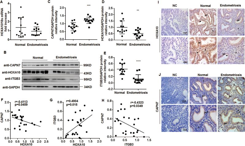
Fig. 7. Aberrant overexpression of CAPN7 in the endometrium of women with ENDO.
a Timed mid-secretory endometrial biopsies from healthy controls (n = 12) and infertile women with ENDO (n = 12) were analyzed for the mRNA expression of HOXA10 by qRT-PCR analysis. b Timed mid-secretory endometrial biopsies from healthy controls (n = 12) and infertile women with ENDO (n = 12) were analyzed for HOXA10 and CAPN7 protein expression using western blot analysis. c The intensities of CAPN7 signals were quantified from the 12 samples and normalized to GAPDH. ***p < 0.001. d The intensities of HOXA10 signals were quantified from the 12 samples and normalized to GAPDH. **p < 0.01 vs control. e The intensities of ITGB3 signals were quantified from the 12 samples and normalized to GAPDH. ****p < 0.0001 vs control. f Correlation between CAPN7 and HOXA10 expression in endometrial samples of women with or without ENDO (r = -0.4113, p = 0.0459). g Correlation between ITGB3 and HOXA10 expression in endometrial samples of women with or without ENDO (r = 0.4904, p = 0.015). h Correlation between CAPN7 and ITGB3 expression in endometrial samples of women with or without ENDO (r = -0.4323, p = 0.0349). i Timed mid-secretory endometrial biopsies from healthy control (n = 3) and infertile women with ENDO (n = 3) were analyzed using immunohistochemistry (IHC). Goat IgG was used as a negative control. Arrows show the increased HOXA10 conjugates in the glandular epithelium. j Timed mid-secretory endometrial biopsies from healthy control (n = 3) and infertile women with ENDO (n = 3) were analyzed using immunohistochemistry (IHC). Rabbit IgG was used as a negative control. Arrows show the increased CAPN7 conjugates in the glandular epithelium

