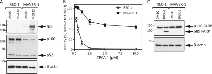
Fig. 1. REC-1 and MAVER-1 cells show distinct dependencies on classical NFκB signaling.
a MCL cell lines were treated with MG132 (20 μM) for 8 h, and whole cell lysates were analyzed by Western blot. b REC-1 and MAVER-1 cells were incubated with increasing concentrations of TPCA-1 for 48 h, and viability was determined via MTT assay. c Whole cell lysates of the MCL cell lines were prepared after treatment with TPCA-1 (5 μM) for 24 h and were subsequently analyzed by Western blot

