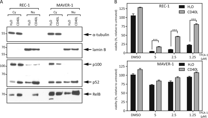
Fig. 2. CD40L rescues IKK2 inhibition-mediated toxicity by activating the alternative NFκB pathway.
a REC-1 and MAVER-1 cells were stimulated with CD40L (100 ng/ml) or H2O as a control for 18 h, and cytoplasmic (Cy) and nuclear (Nu) protein fractions were analyzed by Western blot. α-Tubulin and lamin B served as controls for the purity of the individual cell compartment fractions. b MCL cell lines were stimulated with CD40L (100 ng/ml) or H2O as a control overnight followed by TPCA-1 treatment at the indicated concentration. After an additional 24 h, viability was determined by MTT assay (*p < 0.001; **p < 0.0001; ***p < 0.00001)

