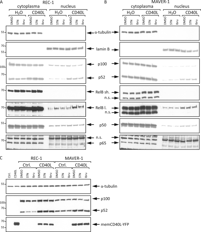
Fig. 3. CD40L activates alternative NFκB signaling independently of the BCR pathway.
The MCL cell lines REC-1 a and MAVER-1 b were preincubated with CD40L (100 ng/ml) or H2O for 18 h and subsequently treated with either sotrastaurin (STN, 3 μM), ibrutinib (Ibru, 400 nM) or DMSO as a control for an additional 24 h. Cytoplasmic and nuclear protein extracts were analyzed by Western blot for the indicated proteins; “sh.” stands for a short and “l.” for a long exposure time for RelB detection. c L-929 cells were transfected with a CD40L expression plasmid or a control plasmid and co-cultured with REC-1 and MAVER-1 cells for 18 h. Subsequently, the cells were treated with sotrastaurin (3 µM), ibrutinib (400 nM) or DMSO for an additional 24 h, and whole cell lysates were analyzed by Western blot

