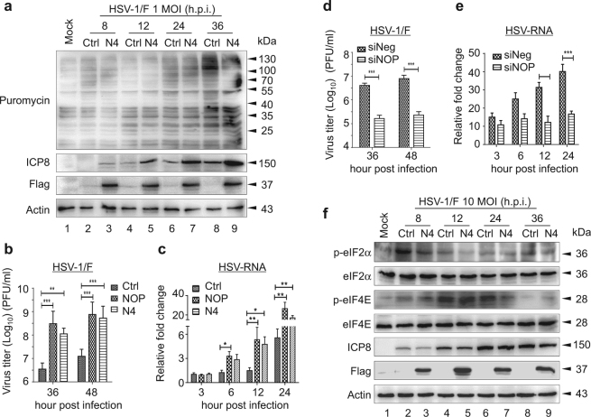
Fig. 2. NOP53 improves viral protein synthesis of HSV-1/F.
a HeLa cells transfected with control plasmid (lanes 2, 4, 6, 8) or Flag-tagged N4 (lanes 3, 5, 7, 9) were mock-infected (lane 1) or infected with HSV-1/F at 1 MOI for indicated times (lanes 2–9). Cells were metabolically pulse-chase labeled with puromycin for 1 h prior to harvest. Cell lysates were analyzed by Western blotting with the indicated antibodies. De novo protein synthesis was assessed using anti-puromycin antibody 12D10. b HeLa cells were transfected with control plasmid, Flag-tagged NOP53, or Flag-tagged N4. The cells were infected with HSV-1/F at 0.1 MOI, and viral yields were determined 36 or 48 h.p.i. by plaque assay. These experiments were performed two times with three replicates in each experiment. Values represent means with SD. **p ≤ 0.01; ***p ≤ 0.001. c HeLa cells seeded in 24-well plate were transfected with control plasmid, Flag-tagged NOP53 or Flag-tagged N4 (500 ng each), and then infected with HSV-1/F at 0.1 MOI for the indicated times. Total RNAs were isolated; expression levels of HSV-ICP8 mRNA were quantified by qRT-PCR and normalized with glyceraldehyde-3-phosphate dehydrogenase (GAPDH). These experiments were performed two times with three replicates in each experiment. Values represent means with SD. *p ≤ 0.05; **p ≤ 0.01; ***p ≤ 0.001. d HeLa cells were transfected with siNOP or siNeg followed by infection with HSV-1/F at 0.1 MOI, and viral yields were determined 36 or 48 h.p.i. by plaque assay. These experiments were performed two times with three replicates in each experiment. Values represent means with SD. ***p ≤ 0.001. e HeLa cells seeded in 24-well plate were transfected with siNOP or siNeg (20 pmol each), and then infected with HSV-1/F at 0.1 MOI for the indicated times, and assessed as described in Fig. 3c. Values represent means of triplicates with SD. **p ≤ 0.01; ***p ≤ 0.001. f HeLa cells transfected with control plasmid (lanes 2, 4, 6, 8) or Flag-tagged N4 (lanes 3, 5, 7, 9) were mock-infected (lane 1) or infected with HSV-1/F at 10 MOI for indicated times (lanes 2–9). Cell lysates were then analyzed by Western blotting using antibodies directed against phospho-eIF2α (p-eIF2α), eIF2α, phospho-eIF4E (p-eIF4E), eIF4E, HSV-ICP8, Flag, and actin

