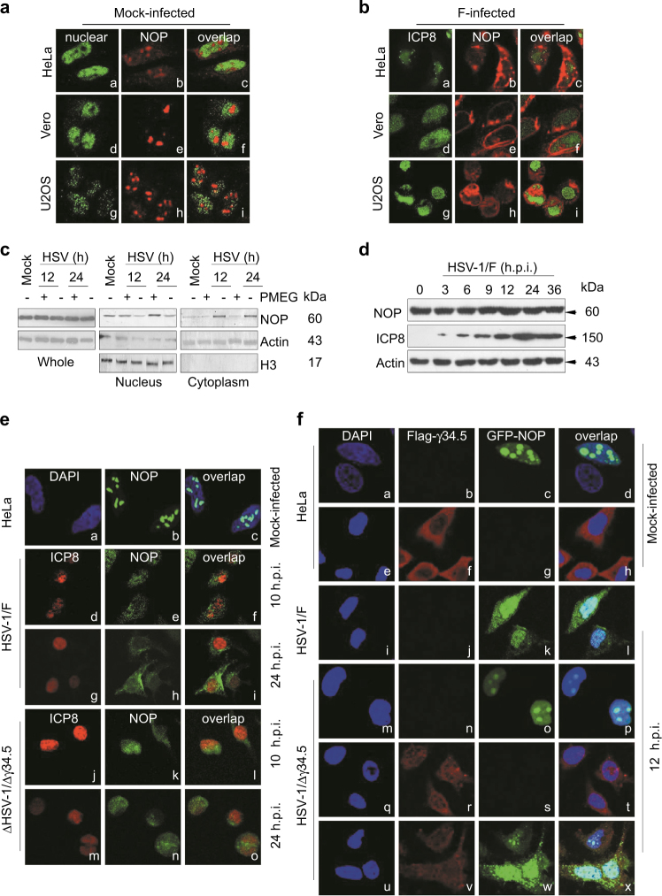
Fig. 4. γ34.5 induces the cytoplasmic translocation of NOP53.
a, b HeLa, Vero, or U2OS cells grown in 4 well slides were mock-infected a or exposed to 10 MOI of HSV-1/F b. At 12 h.p.i., cells were fixed and then reacted with antibodies to NOP53 (b, e, h), ICP8 (a, d, g for panel b), PDCD4 (a, d, g for panel a), and overlap (c, f, i). c HeLa cells were mock-infected or infected with HSV-1/F at 10 MOI for 12 or 24 h, in the presence of PMEG (20 μg/mL) or vehicle control DMSO. The cells were harvested and nuclei and cytoplasm were then isolated and analyzed by Western blotting with an antibody against NOP53. Actin and H3 were used as loading controls for separated cytoplasmic and nuclear proteins, respectively. d HeLa cells were infected with HSV-1/F at 1 MOI for 0, 3, 6, 9, 12, 24, or 36 h, cell lysates were prepared, and NOP53 and HSV-ICP8 were measured by Western blotting with specific antibodies, with actin as a control. e HeLa cells were mock-infected (a–c) or infected with HSV-1/F (d–i), or Δγ34.5 (j–o) at 10 MOI. Cells were fixed and then reacted with antibodies to NOP53 (b, e, h, k, n), HSV-ICP8 (d, g, j, m), and overlap (c, f, i, l, o). Nuclei were stained with DAPI (a). f HeLa cells were transfected with GFP-tagged NOP53 (c, k, o) or Flag-tagged γ34.5 (f, r), or co-transfected with Flag-tagged γ34.5 and GFP-tagged NOP53 (v, w). The cells were then mock-infected (a–h) or infected with either HSV-1/F (i–l) or Δγ34.5 (m–x) at 10 MOI. Cells were fixed and then reacted with antibodies to Flag (b, f, j, n, r, v), GFP (c, g, k, o, s, w), and overlap (d, h, l, p, t, x). Nuclei were stained with DAPI

