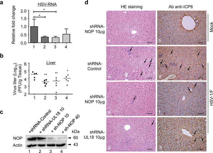
Fig. 8. NOP53 knockdown reduces HSV-1/F growth and pathogenesis in mice.
a BALB/c mice were injected i.p. with control shRNA (lane 1), UL18-specific shRNA (lane 2), NOP53-specific shRNA (lane 3) (10 μg each), or NOP53-specific shRNA (40 μg, lane 4) for 24 h. The mice were then infected with HSV-1/F (107 PFU/ mice). Mice were killed 5 days later and the livers were harvested, total RNAs were isolated. Expression levels of HSV-ICP8 mRNA were quantified by qRT-PCR and normalized with GAPDH. Values represent means of triplicates with SD. *p < 0.05. b Under the same experimental condition, mice livers (n = 5) were homogenized in media and quantified for viral yields by plaque assay, and were presented as log10 PFU/ml. Data are representative of two independent experiments, *p < 0.05; **p < 0.01. c Efficiency of knockdown was analyzed by Western blotting with specific antibody to NOP53, with actin as a control. d Representative images of H&E staining (left) and HSV-ICP8 (right) in liver from mice infected with HSV-1/F. Arrows in left panel indicate eosinophilic, inflammatory cell infiltration, necrotic cells with pyknotic nuclei. Scale bars, 50 μm

