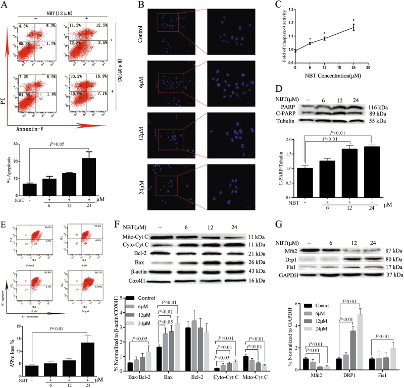
Fig. 4. NBT induced MCF-7 cell apoptosis through the mitochondrial pathway.
a MCF-7 cells were treated with various concentrations of NBT for 24 h, and apoptosis was examined by using annexin V with PI staining with flow cytometry. b Hoechst 33258 staining assay showed that NBT at doses of 6, 12, and 24 μM induced chromatin shrinking of MCF-7 cells (×200, scale bars: 100 μm; ×400, scale bars: 50 μm). c Treatment of 6, 12, and 24 μM NBT influenced caspase-9 activity in MCF-7 cells. d Total cellular extract of MCF-7 cells treated with 6, 12, and 24 μM NBT was prepared and subjected to Western blot by using an antibody against PARP. e JC-1 staining assay determined the ΔΨm in mitochondria of MCF-7 cells treated with 6, 12, and 24 μM NBT. f Total cellular extract, mitochondrial fraction, and cytosol fractions of MCF-7 cells treated with 6, 12, and 24 μM NBT were prepared and subjected to Western blot by using antibodies against Bcl-2, Bax, and Cyt C (mitochondrial and cytosol fraction). g Total cellular extract of MCF-7 cells treated with 6, 12, and 24 μM NBT was prepared and subjected to Western blot by using antibodies against DRP1, Fis1, and Mfn2. Data represent mean ± SD of three independent experiments (*P < 0.05, vs. control group)

