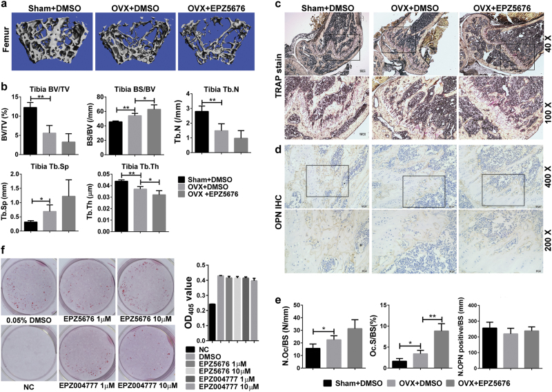
Fig. 3. Inhibition of DOT1L aggravates bone mass reduction in OVX mice.
a OVX mice were sacrificed after 8 weeks of EPZ5676 treatment. Micro-computed tomography images (n = 5) of trabecular bone from femurs in the sham-operated group (Sham), OVX mice, and EPZ5676-treated OVX mice. b Measurements of the ratios of bone volume to total volume (BV/TV), bone surface to bone volume (BS/BV), trabecular number (Tb.N), trabecular spacing (Tb.Sp), and trabecular thickness (Tb.Th) for the indicated mice. c For static histomorphometric analysis, femurs were fixed and embedded in paraffin. The paraffin-embedded bone sections from Sham, OVX, and EPZ004777-treated OVX mice were double-stained with TRAP and hematoxylin (top, low magnification [40 × ] of proximal femoral metaphysis; bottom, high magnification (100 × ) of the black frame in the top panel). d Immunohistochemical staining of osteoblastic markers OPN in femurs from Sham, OVX, and EPZ004777-treated OVX mice. e OC surface/bone surface (Oc.S/BS, %), and OC number/bone surface (N.Oc/BS, N/mm). Number of OPN-positive (N.OPN+) osteoblasts on the bone surface (N.OPN positive/BS, N/mm) was calculated. f MC3T3E1 was induced to differentiate into osteoblasts by treatment with 50 µg/mL l-ascorbic acid and 10 mM β-glycerophosphate in the presence of control or DOT1L inhibitors for 18 days and then fixed with Alizarin S red. Left panel shows Alizarin S red staining. Right panel shows the optical density at 405 nm of the Alizarin S red extract from the left panel. Experimental data are expressed as mean ± standard deviation. *P < 0.05, **P < 0.01, ***P < 0.001, ****P < 0.0001, two-tailed unpaired t-test. OPN: osteopontin.

