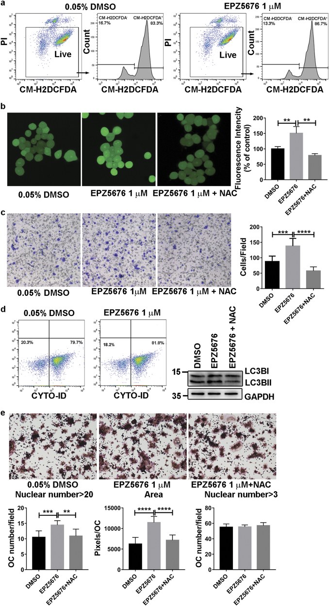
Fig. 7. DOT1L inhibition increases ROS generation, autophagy flux, and migration ability of 40-h pre-OCs.
RAW264.7 cells pretreated with DMSO or the DOT1L inhibitor EPZ5676 (1 µM) were stimulated for indicted time with RANKL. a Flow cytometric analysis of ROS generation in 40-h pre-OCs by PI and CM-H2DCFDA double-staining. PI-negative live cells were gated for CM-H2DCFDA measurement. b Confocal fluorescence microscopy detection of ROS generation in 40-h pre-OCs treated with DMSO, EPZ5676 (1 µM) or EPZ5676 (1 µM) combined with NAC (1 mM). The fluorescence intensity was calculated with ImageJ software (c) Migration analysis of 40-h pre-OCs using transwell migration assays. Cell number per field was counted. Experimental data are expressed as the mean ± standard deviation. d Flow cytometric analysis of autophagy in 40-h pre-OCs using the CYTO-ID autophagy detection kit. e TRAP staining and analysis of OCs treated with DMSO, EPZ5676 (1 µM) or EPZ5676 (1 µM) combined with NAC (1 mM). ***P < 0.001, two-tailed unpaired t-test, compared with DMSO treatment. ROS: reactive oxygen species, DMSO: dimethyl sulfoxide, CM-H2DCFDA: chloromethyl derivative of 2’,7’-dichlorodihydrofluorescein diacetate, PI: propidium iodide.

