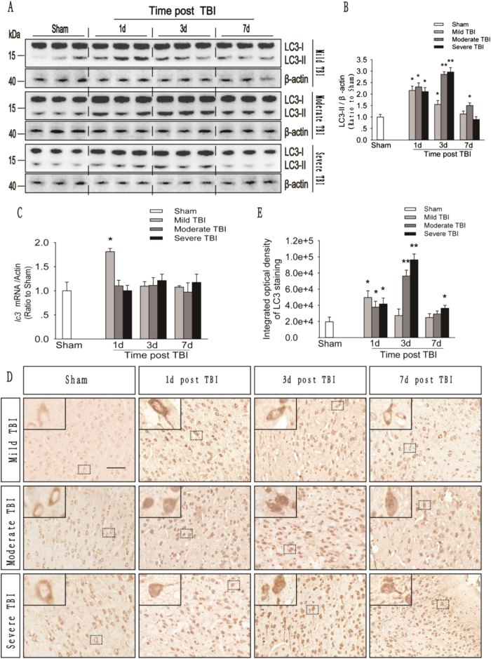
Fig. 1. The number of autophagosomes in the injured cortex increases after mild, moderate and severe TBI.
a Western blot analysis of the levels of the autophagy-related protein LC3 in cortical tissue lysates obtained from sham and TBI mice 1, 3, and 7 days after injury. b The LC3-II levels shown in (a) are quantified and normalized to the β-actin. The data shown are presented as means ± SEM, n = 5–6, *P < 0.05 and **P < 0.01. c Relative mRNA levels (qPCR) of lc3 in the sham and injured mouse cortex. The results are normalized to β-actin levels. The data are presented as means ± SEM, n = 3, *P < 0.05 compared to the sham group. d Images of cortical brain sections obtained from sham and TBI mice. Sections were stained using an anti-LC3 antibody. Scale bar = 50 μm. e Integrated optical density analysis of the LC3 data shown in (d). The data are presented as means ± SEM, n = 5–6, *P < 0.05, **P < 0.01 compared to the sham group

